Image-guided planning for interventional radiology

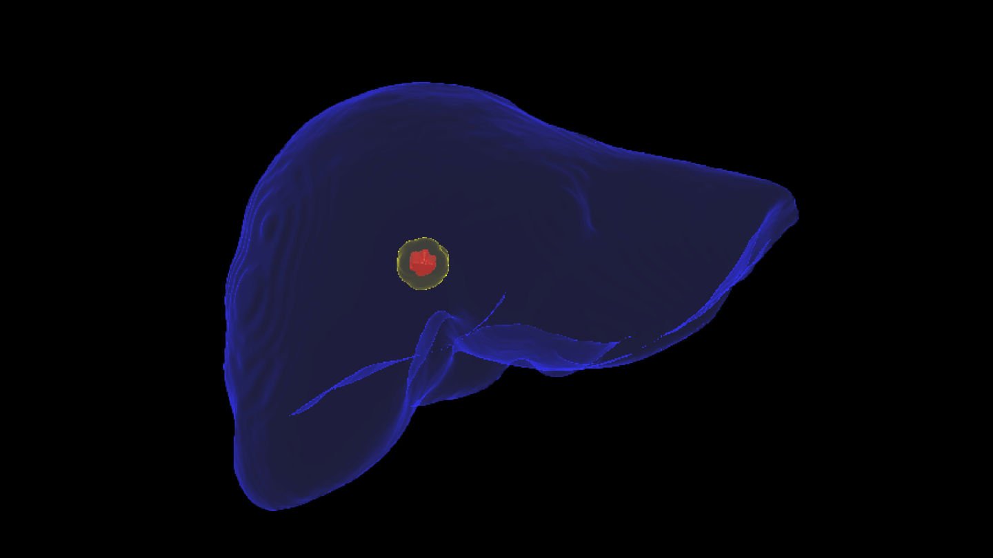
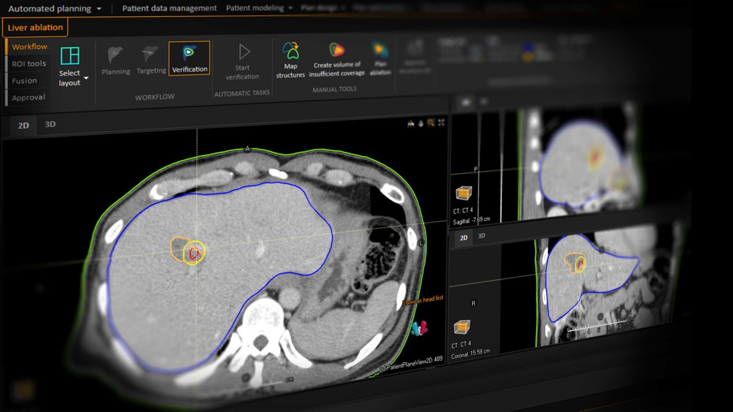
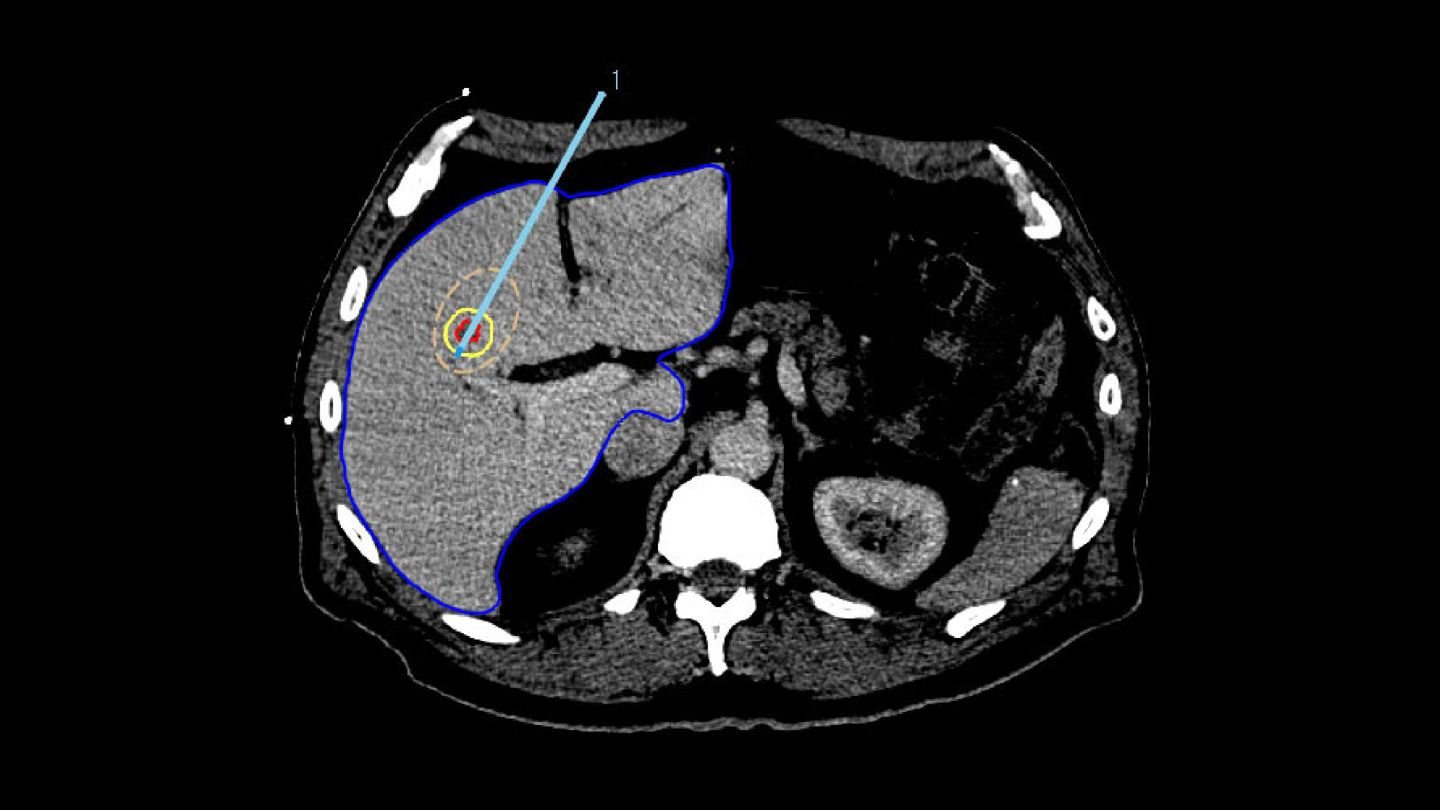
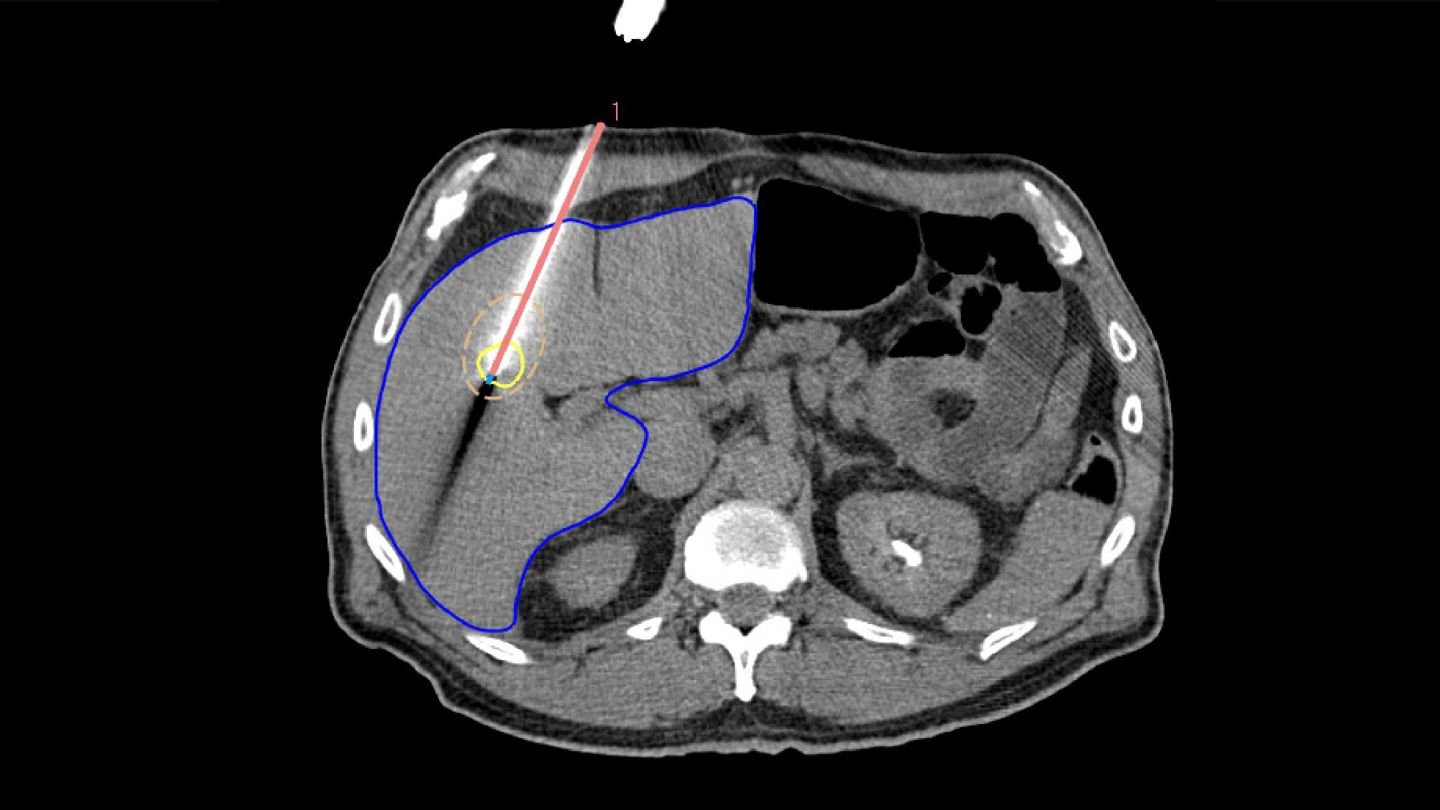
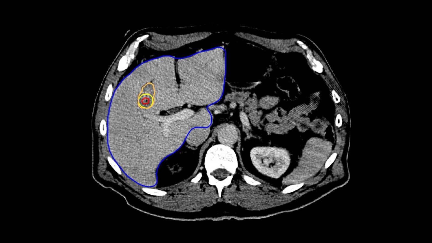
RaySearch takes another step forward in defining comprehensive cancer care by introducing the Image-guided Liver Ablation module*. With the existing tools in RaySearch products, integrating liver ablation was a natural fit, further expanding the range of supported cancer treatment techniques.

All of this is in pursuit of the same comprehensive goal: raising the standard of patient care.


*The module is in an ongoing development phase.

Why liver ablation?

As a treatment technique, liver ablation offers numerous benefits that enhance the patient experience, such as:

  • Shorter procedure times (2 to 3 hours)
  • Fast recovery (patients typically leave just a few hours after the procedure)
  • Minimal side effects, and more.

Liver ablation workflow

A typical liver ablation procedure involves three main steps: planning, targeting, and verification.

The procedure begins with the planning phase, during which the physician decides where to place the applicator. Once the location is determined, the applicator is inserted into the patient during the targeting step.

Finally, to assess whether the desired margin was achieved, verification is performed on a post-treatment image.

RAYSEARCH IMPLEMENTATION

All three of liver ablation procedure steps are seamlessly integrated into our software, giving you full access to an advanced planning toolkit that RaySearch has been developing over many years.

1. PLANNING


The Liver Ablation module offers tools for automatic segmentation of relevant structures. Combined with the ability to place virtual applicators and estimate the ablation zone, our software assists users to determine the optimal number of applicators and their precise placement.

2. TARGETING


To ensure accurate applicator placement before treatment, the segmented tumors from the planning phase are mapped using deformable registration, allowing for a visualization of the anticipated tumor position. Additionally, the ablation zone can be estimated based on the applicator parameters, such as its current position, selected power, and ablation duration.

3. VERIFICATION


For treatment verification, both the minimal ablative margin calculation and visual inspection based on deformable mapped tumors are provided in the Liver Ablation module.

In Paulucci et al., it was shown that using biomechanical deformable registration, the local disease progression risk decreased to less than 1% with an intraprocedurally confirmed minimum ablative margin greater than 4.6 mm.

Why liver ablation in RaySearch?

Over the decades, RaySearch has developed a powerful suite of precise and versatile treatment planning tools.

Advanced features such as deep learning segmentation* and deformable image registration, while seamlessly integrated into RaySearch's software, elevate liver ablation planning to a greater level, making it more predictable and precise.

Read more about how deep learning segmentation brings more consistency to cancer treatment here

*Subject to regulatory clearance in some markets.


DEVELOPING IMAGE-GUIDED LIVER ABLATION


Development began in 2019 through collaboration with The University of Texas MD Anderson Cancer Center, with in-house written scripts for liver ablation treatments performed at MD Anderson. Since then, these scripts have steadily advanced and refined, gradually being integrated into RaySearch’s software.
 
Today, the Liver Ablation module is closer than ever to becoming a complete clinical solution, bringing real advancements in liver ablation treatment to those in need.