A COMPREHENSIVE TOOLBOX FOR PHYSICIANS
Everything physicians need to efficiently represent the patient anatomy and easily compare and approve plans.

Everything physicians need to efficiently represent the patient anatomy and easily compare and approve plans.
RayStation provides you with fast and user-friendly tools to efficiently and accurately contour the patient in the treatment planning process. Contouring OAR. It includes a comprehensive toolset ranging from manual tools to high-end semi-automatic and fully automatic contouring tools. Discover some of our customers’ favorite tools below.
Smart brush and smart interpolation
The image-guided smart brush and smart interpolation facilitate contouring by snapping to image features and can help delineate organs and targets using only a few contours. See video
Structure templates
Structure templates allow you to create ROIs with predefined name, type, and color which allows for consistently labeled structure sets according to the clinics’ protocol. Structure templates may also include geometries including density overrides for quick retrieval of support structures.
ROI algebra
With ROI algebra, you can create derived ROIs using Boolean expressions and margins. A derived ROI remembers its expression and will automatically detect when it needs to be updated. Derived ROIs also goes into structure templates, saving you the time of specifying complex expressions. See video
Model-based segmentation (MBS) delineates organs automatically using statistical shape models for different body sites. The adaptation to new image data utilizes a combination of greyscale gradients and shape statistics.
Multi-atlas-based segmentation (MABS) lets you use multiple atlas templates to automatically contour the patient. It’s fast and easy to build your own atlas templates with both anatomical and derived structures. In addition, multiple atlases can be fused for increased accuracy.
RayStation uses in-house deformable registration algorithms to propagate contours from multiple datasets to the anatomy you wish to contour. Advanced automation makes the process fast, simple and accurate.
Existing data from the clinic database is used to create templates with multiple image sets – atlases. The geometries contained in each atlas can be manually contoured or generated with MBS.
New image data is segmented by locating the best-matching atlases through rigid image registration. For all matching atlases, a deformable registration is computed and the structures are deformed onto the new image set.
Segmentation results are merged to one result using a fusion algorithm. If MBS regions of interest are available in the template, they may be automatically adapted.
“The automatic segmentation is a useful tool for handling the technical complexity of head and neck IMRT as well as the daily management of an important patient flow. In head-and-neck cancer, we have gained significantly for the contouring of healthy organs. We will soon begin using multi-atlas-based segmentation in our clinical routine for lymph node delineation.”
Xavier Liem
Radiation Oncologist, Centre Oscar Lambret, Lille, France

The UK’s Royal Marsden Hospital carried out an assessment of fully-automated atlas-based segmentation for a novel oral mucosal surface organ-at-risk. The center found negligible differences when comparing results for manual and fully automated segmentation, concluding that atlas-based segmentation is a suitable approach for this application. Read the study here.
Royal Marsden Hospital
NHS Foundation Trust

“With more complex RT, delineation of organs at risk (OAR) has become a mandatory but also very time-consuming task for highly skilled staff. Multi-atlas segmentation in RayStation is a highly promising approach that can minimize the human resources needed for delineating OAR.”
Martin Berg
Head of Medical Physics Clinical Practice, Vejle Hospital, Denmark

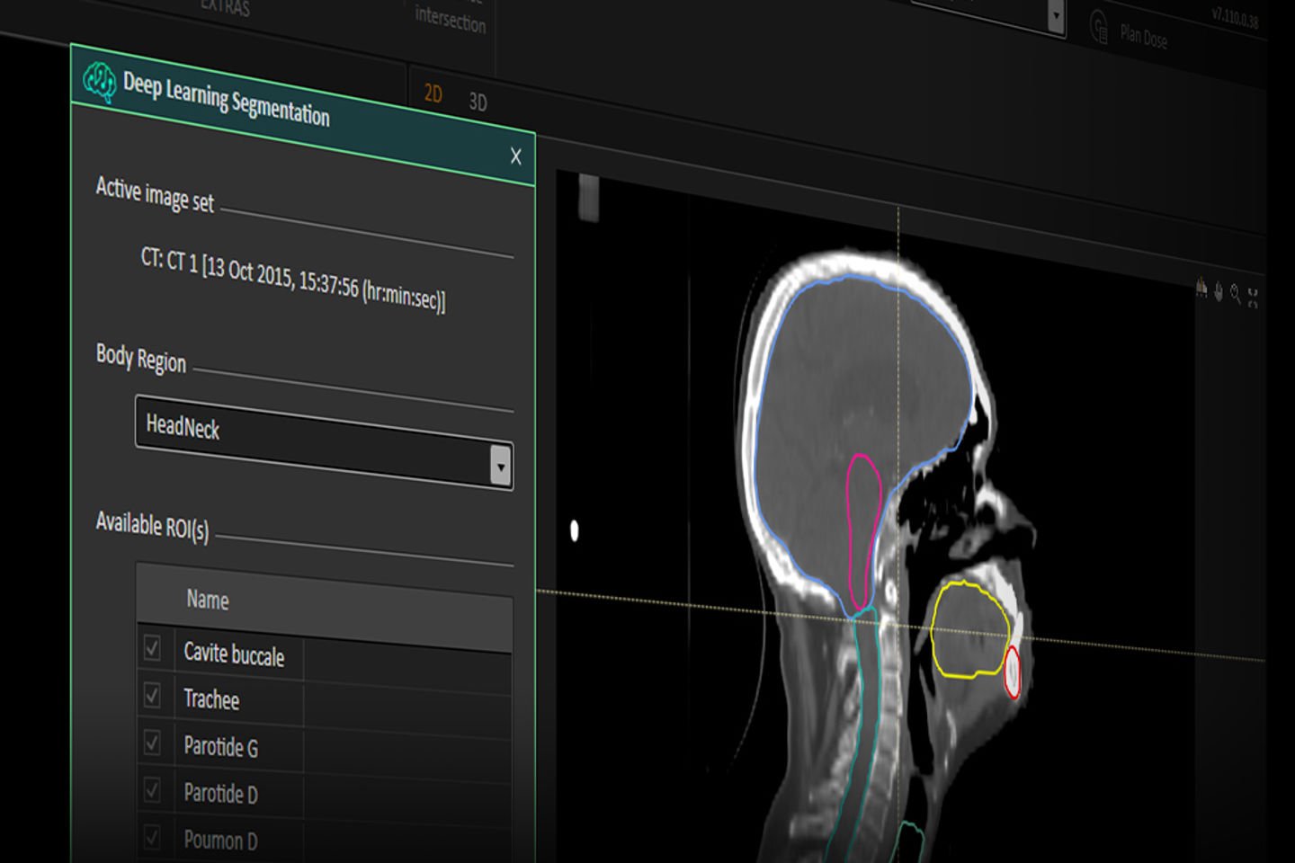
Auto-segmentation of organs at risk in RayStation is set to improve with the upcoming introduction of deep learning segmentation*. The algorithm uses models that have been trained and evaluated on clinical data for different body sites. The GPU-powered algorithm is fast and produces consistently excellent segmentation results.
Register for the upcoming webinar to learn more.
* Subject to regulatory clearance in some markets.

Physicians can easily evaluate, compare and approve treatment plans.
The module is a comprehensive toolbox for evaluation and comparison of treatment plans and plan approval. From a number of predefined layouts, dose distribution, dose statistics, clinical goals and dose volume histograms of up to three different plans can be simultaneously compared. Dose can directly be computed on additional image sets and summed up using the deformable registration provided in rayDeformable. For evaluation of robustness dose can also be computed for a density perturbation or isocenter shift.
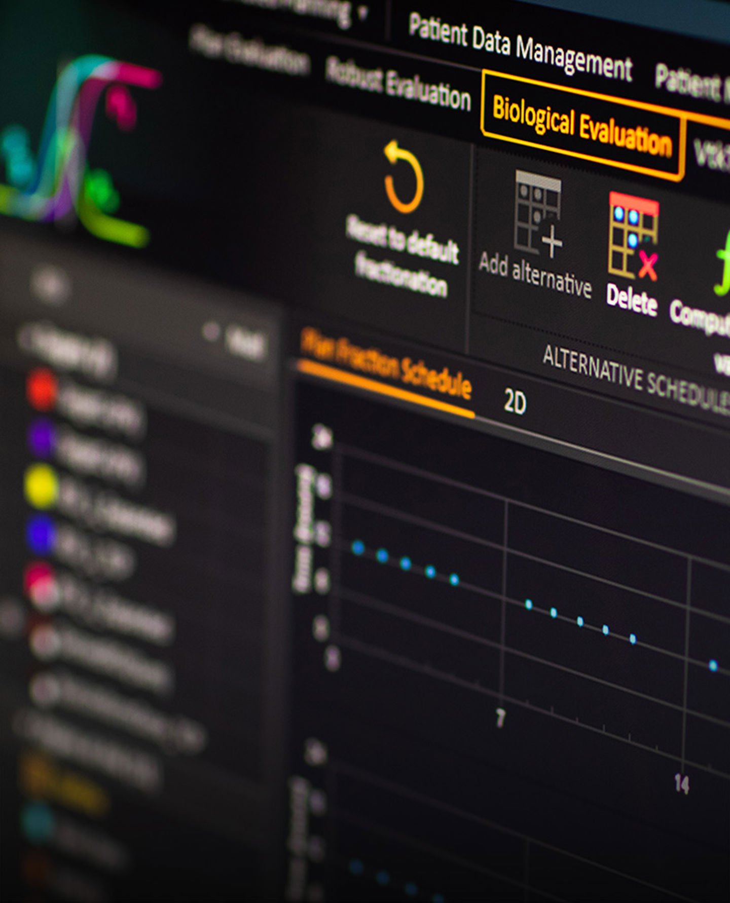
The use of radiobiological response models brings the planning process closer to the intent to create a plan with the highest possible probability of curing the patient while keeping the risk of complications to the surrounding healthy tissue as low as possible. The philosophy is to use biological models to reveal effects that are difficult to understand from the dose distribution alone.
There are tools for both biological optimization and evaluation. Optimization tools enable direct optimization on the biological indices in combination with physical dose indices. This makes it possible to configure optimization problems that more closely describe the clinical intentions, such as minimizing normal tissue complication probabilities subject to a guaranteed homogenous target dose within a specified standard deviation.
Evaluation tools include Tumor Control Probability (TCP) and Normal Tissue Complication Probability (NTCP) models which can be combined with tissue repair and tumor growth models. The biological tools are integrated into the plan evaluation module. The physical properties of the existing plan can thus be easily investigated if unexpected differences in the biological response are observed between different plans. In addition, there is a biological evaluation module dedicated to the exploration of the biological effects of altering the fractionation schedule of a single treatment plan.
